April 19, 2021
Analysis of Surgical Specimens of Coronavirus Disease 2019 (COVID-19) Pneumonia
~Toward the elucidation of the mechanism of sequelae
This result was presented at the General Thoracic and Cardiovascular Surgery on April 3, 2021, and the efforts of the research team including this case will be presented at the 38th Annual Meeting of the Japanese Association for Chest Surgery on May 21.
Key Points
- The lung resection was safely performed on a patient following treatment for COVID-19 pneumonia.
- This is the first study reporting the histological features of the lung specimen after treatment of COVID-19 pneumonia.
- The study is expected to clarify the mechanism of sequelae following COVID-19 pneumonia treatment.

The coronavirus disease 2019 (COVID-19) has many unsolved problems, such as sequelae and a high mortality rate of perioperative infection. A research group led by Professor Akira Iyoda and Assistant Professor Takashi Sakai of the Division of Chest Surgery, Department of Surgery, Toho University Faculty of Medicine have succeeded in safely performing lobectomy in patients after treatment for COVID-19 pneumonia by setting an appropriate waiting period. Following histological and immunological analyses of the resected lung specimen, they found that even after a long period since infection, the lungs showed specific changes due to pneumonia, such as fibrosis, inflammatory cells infiltration, and intravascular hemorrhagic thrombosis. This finding is expected to elucidate the mechanism of sequelae after treatment for COVID-19 pneumonia, the cause of which is currently unknown.
This result was presented at the General Thoracic and Cardiovascular Surgery on April 3, 2021, and the efforts of the research team including this case will be presented at the 38th Annual Meeting of the Japanese Association for Chest Surgery on May 21.
Key Points
- The lung resection was safely performed on a patient following treatment for COVID-19 pneumonia.
- This is the first study reporting the histological features of the lung specimen after treatment of COVID-19 pneumonia.
- The study is expected to clarify the mechanism of sequelae following COVID-19 pneumonia treatment.
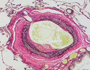
Organized hemorrhagic thrombosis were found in peripheral pulmonary vessels.
Although the pathogenesis and treatment for COVID-19 infection have been gradually clarified, there are still unresolved problems, such as the sequelae after the infection. In addition, a high mortality rate has been reported in COVID-19 infected patients who underwent thoracic surgery. The guidelines recommend the elective surgery or alternative treatment for the patients with thoracic malignancies during the COVID-19 pandemic period, however, there is no evidence and guidelines about the elective surgery. In the present study, the research group succeeded in performing lobectomy safely in lung cancer patients after treatment for COVID-19 pneumonia by setting an appropriate waiting period. By analyzing the resected lung histologically and immunologically, we found that while the operation was safe and non-infectious, the characteristic histopathological images such as intravascular hemorrhagic thrombosis and fibrosis remained for a long time following COVID-19 pneumonia treatment. An accumulation of such cases will provide guidelines for setting the appropriate treatment timing for patients requiring thoracic surgery, besides being useful in elucidating the mechanism of sequelae.
- Takashi Sakai (Assistant Professor, Division of Chest Surgery, Department of Surgery, Toho University Faculty of Medicine)
- Akira Iyoda (Professor, Division of Chest Surgery, Department of Surgery, Toho University Faculty of Medicine)
- Kazuhiro Tateda (Professor, Department of Microbiology and Infectious Diseases, Toho University Faculty of Medicine)
- Sakae Homma (Professor, Department of Advanced and Integrated Interstitial Lung Diseases Research, Toho University Faculty of Medicine)
- Kazuma Kishi (Professor, Division of Respiratory Medicine, Department of Internal Medicine, Toho University Faculty of Medicine)
- Shion Miyoshi (Assistant Professor, Division of Respiratory Medicine, Department of Internal Medicine, Toho University Faculty of Medicine)
- Megumi Wakayama (Department of Surgical Pathology, Toho University Faculty of Medicine)
- Kotaro Aoki (Assistant Professor, Department of Microbiology and Infectious Diseases, Toho University Faculty of Medicine)
- Yoko Azuma (Division of Chest Surgery, Department of Surgery, Toho University Faculty of Medicine)
READ MORE RESEARCH NEWS - MEDICINE
Undergraduate Programs
– Medicine
– Pharmaceutical Sciences
– Science
– Nursing
– Health Science
Graduate Programs
–Medicine
–Pharmaceutical Sciences
–Science
–Nursing
RESEARCH
– News
– Guidelines & Policies
– Support Offices
– Facilities
– Security Export Control
Non-Degree Programs
– Clinical Elective Program
– International Physician Observership Program
