This result was presented at the General Thoracic and Cardiovascular Surgery on April 3, 2021, and the efforts of the research team including this case will be presented at the 38th Annual Meeting of the Japanese Association for Chest Surgery on May 21.
Key Points
- The lung resection was safely performed on a patient following treatment for COVID-19 pneumonia.
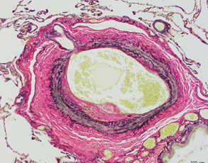
- This is the first study reporting the histological features of the lung specimen after treatment of COVID-19 pneumonia.
- The study is expected to clarify the mechanism of sequelae following COVID-19 pneumonia treatment.
The coronavirus disease 2019 (COVID-19) has many unsolved problems, such as sequelae and a high mortality rate of perioperative infection. A research group led by Professor Akira Iyoda and Assistant Professor Takashi Sakai of the Division of Chest Surgery, Department of Surgery, Toho University Faculty of Medicine have succeeded in safely performing lobectomy in patients after treatment for COVID-19 pneumonia by setting an appropriate waiting period. Following histological and immunological analyses of the resected lung specimen, they found that even after a long period since infection, the lungs showed specific changes due to pneumonia, such as fibrosis, inflammatory cells infiltration, and intravascular hemorrhagic thrombosis. This finding is expected to elucidate the mechanism of sequelae after treatment for COVID-19 pneumonia, the cause of which is currently unknown.
This result was presented at the General Thoracic and Cardiovascular Surgery on April 3, 2021, and the efforts of the research team including this case will be presented at the 38th Annual Meeting of the Japanese Association for Chest Surgery on May 21.
Key Points
- The lung resection was safely performed on a patient following treatment for COVID-19 pneumonia.
- This is the first study reporting the histological features of the lung specimen after treatment of COVID-19 pneumonia.
- The study is expected to clarify the mechanism of sequelae following COVID-19 pneumonia treatment.