TOPICS
DATE
October 1, 2021
SHARE
October 1, 2021

Reorientation of Protein-Bound Water Regulates the Magnetic Compass of Organisms -Electron Transfer Mechanism of Photoreceptor Cryptochromes

Dr. Tatsuya Iwata
A joint research team, including Dr. Tatsuya Iwata of the Toho University Faculty of Pharmaceutical Sciences, has conducted structural analyses of the intermediates in a photoreceptor protein, as a model for an important role in magneto-optical sensing in animals, using electron spin resonance, and elucidated the electronic functions inside the proteins that endow them with magnetic compass properties. For the first time, their results reveal the role of protein-bound water motility in the magnetic compass, which is thought to be the origin of light sensitivity and geomagnetism exhibited by various organisms, such as migratory birds. This finding will broaden our understanding of signal transduction mechanisms involved in magneto-optical sensing by living organisms and is expected be applicable in the development of biomolecule-based weak magnetic sensors. The results of this study were published in the British scientific journal “Communications Chemistry” on September 30, 2021.
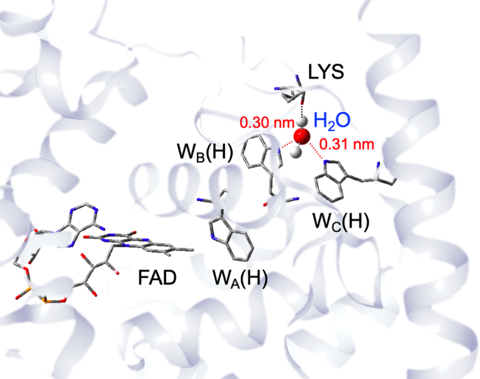
Photoreceptor proteins called cryptochromes are found in various organisms, including migratory birds and plants. Cryptochromes contain FAD, a blue-light-absorbing dye. Blue-light excitation of this dye causes a chemical reaction that progressively withdraws electrons from neighboring tryptophan residues (WA(H), WB(H), WC(H)), resulting in the long-range charge-separated state FAD —-WC(H)+– (Figure 1). This state exists as a magnetic radical pair with a fixed lifetime, but the reaction yield is affected by the electron spin state and the direction and intensity of the external magnetic field. Therefore, a hypothesis has been proposed that migratory birds sense the direction of the geomagnetic field based on the quantum mechanical effect (magnetic compass) of the magnetism produced by this long-range charge-separated state.
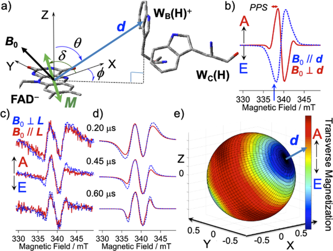
However, the FAD—-WB(H)+– charge-separated state, which is generated in the first stage before the long-range FAD—-WC(H)+– charge-separated state is generated in the third and later stages, is short-lived, and details of its three-dimensional structure and mobility have not been determined. Previously, Prof. Yasuhiro Kobori’s (Department of Chemistry, Graduate School of Science, Kobe University, Molecular Photoscience Research Center, Kobe University) group has used time-resolved electron spin resonance to analyze the structures of the initial photo-charge separated states in various systems, such as photosystem II in plant photosynthesis and organic solar cells. In particular, they have developed the first spin-polarization imaging technique, in which the anisotropy of electron spin polarization generated in the intermediate spin state is resolved in the spatial direction of the external magnetic field and its intensity is projected, thus enabling the visualization of the three-dimensional arrangement of photoreaction intermediates as three-dimensional images. This method is expected to reveal the three-dimensional structures of photoreaction intermediates of cryptochromes, and contribute to elucidating the entire magnetic compass mechanism by enabling the investigation of electron orbitals and molecular motion of the intermediates.

Fig. 1: Water binding between WB(H) and WC(H) forming an electron-tunneling route in chryptochrome.
X-ray structure of the animal-like cryptochrome of Chlamydomonas reinhardtii (PDB code:5ZM0) with the Trp triad comprising WA(H), WB(H) and WC(H).

Fig. 2: Molecular conformation analyses of the secondary RP state.
a Geometry setting of the secondary CS state with the transition dipole moment (M) lying in the FAD aromatic X-Y plane with δ = 65°.<b.b The singlet precursor SCRP spectra computed for B0 // d (dashed line) and for B0 d (solid line). c Magnetophotoselection (MPS) effects of the TREPR spectra for the delay times of td = 0.20, 0.45 and 0.60 μs at 120 K with B0 L (dashed line) and B0 // L (solid line). d Computed EPR spectra of the SCRP for the B0 L (dashed line) and for B0 // L (solid line) with applying D = −0.90 mT, θ = 58°, and ϕ = −65°. J = 1.45, 0.55 and 0.40 mT were applied for td = 0.20, 0.45, and 0.60 μs, respectively. T23 = 0.25 μs was utilized as the relaxation time constant between |2> and |3> by J-modulation, see Fig. 2d. e Mapping of the electron spin polarization (ESP) obtained by distributing the transverse magnetization (EPR intensities at B0 = 337.50 mT shown by blue arrow in b as the color map to the B0 space directions from the SCRP spectra at td = 0.45 μs, demonstrating that the d vector directs to WB(H) in the reference X-Y-Z coordinate system in a) of the protein.

Authors:
Misato Hamada, Department of Chemistry, Graduate School of Science, Kobe University
Tatsuya Iwata, Department of Pharmaceutical Sciences, Toho University
Masaaki Fuki, Department of Chemistry, Graduate School of Science, Kobe University
Molecular Photoscience Research Center, Kobe University
Hideki Kandori, Department of Life Science and Applied Chemistry, Nagoya Institute of Technology, OptoBioTechnology Research Center, Nagoya Institute of Technology,
Stefan Weber, Institute of Physical Chemistry, Albert-Ludwigs-Universität Freiburg
Yasuhiro Kobori, Department of Chemistry, Graduate School of Science, Kobe University, Molecular Photoscience Research Center, Kobe University

READ MORE RESEARCH NEWS - Pharmaceutical Sciences

@ Toho University