The research group led by Associate Professor Masayoshi Tokita (Department of Biology, Faculty of Science) explored the molecular basis generating the diversity of amniote skull morphology, using embryos of several amniote species as materials. The study suggests that the diversity of amniote skull morphology may be brought about by spatiotemporal differences in the expression of three osteogenic genes Msx2, Runx2, and Sp7.The finding provides a basis for understanding how skull morphology has diversified in amniotes, the first vertebrate lineage fully move to land, including human. Furthermore, the finding may provide hints for the development of treatments for congenital diseases that cause abnormalities in human skull morphology.
The finding of this research was reported by the American scientific journal Science Advances on November 15, 2023.
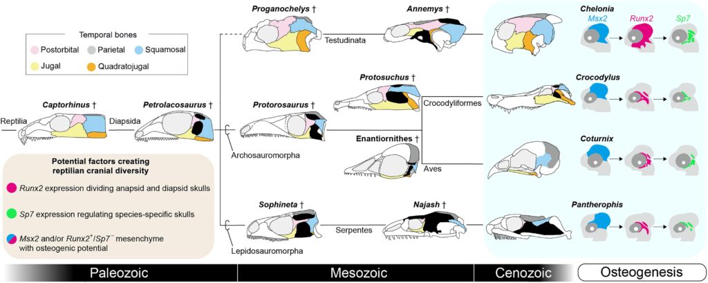
Evolutionary history of temporal skull morphology in Reptilia and developmental insights into cranial diversification.
Simplified phylogeny of the clade Reptilia including fossil (†) and living species. The skull illustration of Enantiornithes is represented by IVPP V12707 (36). Living species analyzed in this study are highlighted by a light blue background. The five temporal dermal bones are shown in distinct colors. Expression domains of Msx2 (blue), Runx2 (magenta), and Sp7 (green) are shown on the embryonic head illustrations of each extant species. Widespread expression of Msx2 is shared in the temporal mesenchyme in all species examined. Runx2 expression is broad in turtles but restricted in other species, likely reflecting anapsid and diapsid skull morphologies. Species-specific Sp7 expression within Msx2– and/or Runx2-positive temporal mesenchyme may contribute to the diversification of cranial morphology. During reptilian evolution, the reappearance of temporal arches, the closure of temporal fenestrations, and the roofing of temporal emarginations by bones have occurred multiple times, resulting in diversification of cranial morphology. This may be triggered by repeated activation of Sp7 in Msx2– and/or Runx2-positive mesenchyme with osteogenic potential.
Journal
Science Advances November 15, 2023 issue
Title
Turtle skull development unveils a molecular basis for amniote cranial diversity
Authors
Hiromu Sato, Noritaka Adachi, Satomi Kondo, Chiyo Kitayama, and Masayoshi Tokita*
DOI No.
10.1126/sciadv.adi6765