TOPICS
DATE
September 2, 2022
SHARE
September 2, 2022

Development of TiYO for autofluorescence quenching, tissue staining and multiple fluorescent staining

Dr. Yousuke Tsuneoka
A research group consisting of Dr. Yousuke Tsuneoka and Professor Hiromasa Funato of the Department of Anatomy, Toho University Faculty of Medicine, has developed TiYO™, an LED-based fluorescence quenching device for use on tissue specimens. This device can efficiently quench autofluorescence, which is noise in tissues, by significant amounts in a short time. Furthermore, since it can also decolorize tissue stained with fluorescent elementdyes, it is possible to obtain more information from tissue sections than ever before by re-staining a single glass slide multiple times. This device is expected to be applied not only to the development of imaging technology in histology, but also to the detection of valuable specimens in clinical settings. The results were published in Frontiers in Molecular Neuroscience on September 2, 2022.
Key Points:
  • Development of a system capable of quenching autofluorescence that causes noise in tissue specimens.
  • The quenching system is equipped with a vaporization heat cooling system to achieve highly efficient quenching using high-power LEDs and to minimize tissue damage caused by intense light irradiation.
  • Quenching of fluorescent signals from once-stained specimens enables multiple analyses of biological materials from a single slide glass.
Summary
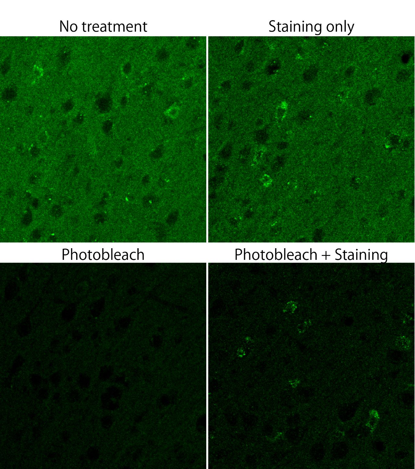
Fluorescent dyes can be used to detect various biological substances in tissues and are indispensable for understanding biological phenomena. However, some substances in living organisms emit fluorescence as noise, which is difficult to distinguish from artificially labeled fluorescence. While recent advances in detection technology have led to higher sensitivity and resolution in fluorescence detection, little progress has been made in dealing with autofluorescence as noise, and new quenching technology has been long awaited. The research group has been developing “quenching technology by light irradiation,” a completely different approach from the reagent-based quenching technology that has been mainly used until now. This new technology has succeeded in quenching autofluorescence practically in a short period of time. By using this device prior to fluorescence staining, it is possible to eliminate only tissue-derived autofluorescence noise without degrading the labeled fluorescence signal. The device can also be used after fluorescence staining to return the sample to its pre-stained state, making it possible to perform fluorescence staining of the same tissue sample multiple times.

Fig. 1 Quenching treatment by TiYO on the mouse brain tissue.

Fig. 2 Comparison of effect of quenching treatment on the fluorescent dyes..

Journal:
Frontiers in Molecular Neuroscience, September 2, 2022 issue

Title:
Fluorescence quenching by high-power LEDs for highly sensitive fluorescence in situ hybridization.

Authors:
Yousuke Tsuneoka*, Yusuke Atsumi, Aki Makanae, Mitsuru Yashiro, Hiromasa Funato*

DOI No:
10.3389/fnmol.2022.976349

READ MORE RESEARCH NEWS - MEDICINE

@ Toho University